NewTom GO 2D/3D CBCT
Advanced 3D imaging. Right here in Spooner.
Advanced 3D Imaging. Right Here in Spooner.
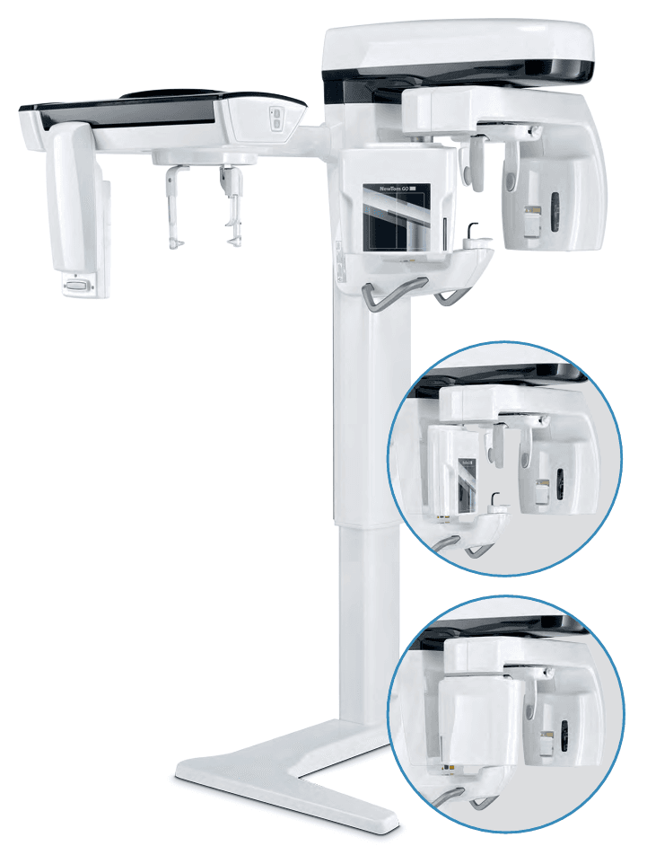
We use the NewTom GO — a state-of-the-art cone beam 3D imaging system that gives us a complete picture of your teeth, jaw, and sinuses in a single, low-dose scan. Better images mean better care.
~10 sec
Average scan time
Low-dose
Less radiation than traditional CT
One visit
No referral needed
3D
Full view of teeth, jaw & sinuses
Why It Matters
What This Means for You
The NewTom GO combines 2D panoramic, 3D cone beam, and cephalometric imaging into one compact system. Here's why that makes a difference in your care.
Minimum X-Ray Dose
SafeBeam™ technology automatically adjusts radiation to your size and the area being scanned — so you're never exposed to more than necessary. ECO protocols reduce dose even further.
Broad Diagnostic Potential
From implant planning and root canals to sinus studies and orthodontic evaluations — one device covers it all, so you don't need to be referred elsewhere for imaging.
Accessible Technology
Guided procedures and smart automatic features make every scan quick and comfortable. The open design accommodates patients in wheelchairs and those with limited mobility.
Maximum Connectivity
Your images can easily be stored, exported, and shared with specialists using industry-standard DICOM formats — making referrals and second opinions seamless.
3D Imaging
Clinical Excellence in Three Dimensions
Using CBCT (Cone Beam Computed Tomography) — a technology NewTom pioneered in dentistry — the GO produces high-definition 3D volumes with just one scan. The field of view ranges from a focused 6×6 cm for detailed endodontic work up to a full 10×10 cm for complete arch views including the sinuses.
The aMAR (autoadaptive Metal Artifact Reduction) algorithm ensures clear images even when metal restorations, amalgam, or implants are present — automatically recognizing metal elements and generating cleaner views.
10×10
centimeters
Complete adult dentition with sinuses. Ideal for full-arch views and third molar assessments.
8×7
centimeters
Complete child dentition. Sized for pediatric patients and orthodontic applications.
6×6
centimeters
Ultra-high resolution local views. Perfect for endodontic and periodontic work.
8×6
centimeters
Localized anatomy — great for sinus lifts and implant site assessment.
10×7
centimeters
Mandibular region analysis. Impacted canine and nerve canal evaluation.
8×10
centimeters
Maxillary sinuses and upper airways with the full superior dental arch.
2D Imaging
Complete Panoramic Vision
The GO delivers sharp, clear panoramic images using a newly developed CMOS sensor and ApT (Autoadaptive Picture Treatment) filters that automatically optimize every image for the specific anatomical region being examined.
The adaptive PAN function captures five focal layers in a single scan — letting us choose the clearest view for your specific diagnostic needs, and the ORTHO mode captures the dental arch orthogonally to eliminate overlap.
Adult & Child Panoramic
Standard panoramic views of dental arches, sinuses, and TMJ. Pediatric mode adapts the field of view and exposure to a child's build, reducing dose.
Orthogonal Panoramic
Captures the dental arch at a right angle to highlight interproximal spaces and root structure without overlap — ideal for detecting hidden decay.
TMJ Imaging
Dedicated trajectories generate four projections in one scan — two lateral and two posteroanterior — with mouth open or closed.
Maxillary Sinus
Specialized focal layer designed for sinus exams, producing both frontal and lateral slices for thorough evaluation.
Bitewing
Collimated interproximal projection for studying dental crowns — a comfortable, low-dose alternative to intraoral bitewings.
Dentition Mode
Focused images limited to the teeth with orthogonal projection and enhanced signal-to-noise ratio. Ideal for periodontal monitoring.
Cephalometric Imaging
Complete CEPH Capabilities
The integrated teleradiographic arm extends our diagnostic range to full cephalometric examinations — critical for orthodontic treatment planning. With dedicated protocols for both adults and children, collimation designed to minimize dose, and scan times as quick as 3.7 seconds.
Longer ear guards for pediatric patients include the skullcap in the scan while reducing thyroid exposure — because extra care for kids is built into the system.
Lateral Cranial Teleradiography
Detailed bone structure views with highlighted soft tissues — essential for cephalometric studies and orthodontic planning.
Frontal Cranial Teleradiography
Frontal projections for scanning for asymmetries and malocclusions to complete treatment planning.
Carpal Teleradiography
Residual growth potential assessment through carpal examination, with a dedicated support for correct scan positioning.
Patient Safety
Minimum Dose, Maximum Quality
NewTom pioneered pulsed emission in dental cone beam imaging over 20 years ago. Every protocol on the GO is designed to get the diagnostic information we need while keeping your exposure as low as possible.
ECO Quality
Ultra-rapid 6.4s scan. Ideal for surgical follow-ups. Minimum effective dose of 9 μSv at 6×6 cm.
Regular Quality
Standard 9.6s scan. Balanced dose and detail for treatment planning and diagnostics.
Best Quality
High-resolution 16.8s scan at 80 μm voxel size. For detailed analysis of micro-structures.
SafeBeam™ Technology
Developed and patented by NewTom, SafeBeam™ automatically adapts the radiation dose to your specific anatomy in the chosen examination area. It controls both the power and intensity of radiation, producing clear, detailed images regardless of bone dimensions and density — without exposing you to anything unnecessary.
ECO PAN — 5 μSv
Ultra-rapid 6.6-second panoramic scan with variable collimation for adults and children. Minimal dose, full diagnostic quality.
ECO CEPH — 3.7 seconds
Minimized cephalometric scan time designed especially for pediatric patients. Longer ear guards protect the thyroid.
Software & Connectivity
Advanced Software, Seamless Sharing
The NewTom NNT software platform handles everything from image acquisition to treatment planning — and connects seamlessly with specialist software, practice management systems, and cloud-based collaboration tools.
NNT 2D & 3D Platform
Certified imaging software for performing, processing, displaying, and sharing all 2D and 3D examinations. Integrated into our daily clinical workflow.
Implant Planning
3D volume processing with realistic implant simulation using extensive libraries. Evaluate anatomy adjacent to implant sites, bone density, and insertion angles.
DICOM & RIS/PACS
Full IHE-compliant connectivity with print, worklist, storage commitment, and query/retrieve. Your images go where they need to go.
Cloud Sharing
Multi-platform cloud workflow for sharing images and plans between implantologists, dentists, and lab technicians — so your care stays coordinated.
Technical Details
Specifications at a Glance
| Specification | Details |
|---|---|
| 3D Resolution | ≥ 6 LP/mm — Voxel size as small as 80 μm |
| 3D Field of View | 6×6 cm up to 10×10 cm (8 FOV options) |
| 3D Scan Times | ECO: 6.4s | Regular: 9.6s | Best: 16.8s |
| 2D Resolution | PAN: 5–6.9 lp/mm (pixel 100–73 μm) | CEPH: 5.6 lp/mm (pixel 89 μm) |
| 2D Panoramic FOV | 210 × 115 mm (adult) | 180 × 100 mm (child) |
| CEPH FOV | 258 × 194 mm |
| PAN Scan Times | ORTHO: 13.7s | Standard: 12.2s | ECO: 6.8s |
| CEPH Scan Times | Regular: 9.9s | ECO: 3.7s |
| Effective Dose (3D) | FOV 10×10: 35–121 μSv | FOV 6×6: 9–40 μSv |
| Effective Dose (2D PAN) | 5–9 μSv |
| 3D Sensor | Amorphous Silicon (CsI) — 16-bit, 65,536 grey levels |
| 2D/CEPH Sensor | CMOS (CsI) — 14-bit, 16,384 grey levels |
| Generator | Constant potential (DC) — 2D: 60–85 kV | 3D: 90 kV pulsed |
| Dose Control | SafeBeam™ automatic exposure adaptation |
| Connectivity | LAN/Ethernet — DICOM 3.0, TWAIN, VDDS, Cloud sharing |
| Patient Positioning | 5-point head support, 3 laser guides, servo-assisted alignment, scout view |
| Accessibility | Wheelchair accessible, two-speed column height adjustment |
See What Better Imaging Looks Like
Have questions about our technology? Ready to schedule an appointment? We'd love to show you what advanced imaging can do for your care.